You wake up on the first morning of your period with a dull, familiar ache in your lower abdomen. As the day unfolds, the sensation may intensify and spread into the lower back, hips, or thighs. Sitting feels uncomfortable, standing offers little relief, and daily plans are quietly rearranged around the pain.
If this is your experience, you are not alone — and it is not a failure of your body.
Menstrual pain is one of the most common gynecological symptoms worldwide, yet many people receive little explanation beyond being told that it is “normal.” Understanding what is happening inside the body can be an important step toward responding to menstrual pain with greater clarity and more effective support (Proctor & Farquhar, 2006; Rajput, 2021).
What Menstrual Pain Is from a Physiological Perspective
Menstrual pain, medically termed dysmenorrhea, results from coordinated uterine contractions that help shed the uterine lining during menstruation. These contractions are regulated by prostaglandins — hormone-like signaling substances that influence the timing, strength, and frequency of uterine muscle activity.
Prostaglandins function as chemical messengers. When their levels are elevated, uterine contractions become stronger and more frequent. This increased contractile activity can temporarily reduce blood flow to the uterine muscle, leading to a short-term decrease in oxygen supply, which is thought to contribute to pain perception (Rosenwaks & Seegar-Jones, 1980).
Menstrual pain may present as:
lower abdominal cramping
pelvic or lower back pain
pain radiating into the hips or thighs
accompanying symptoms such as nausea, headache, dizziness, or fatigue
The intensity and pattern of pain vary widely between individuals and may also change from cycle to cycle (Vijayasri et al., 2023).
Primary and Secondary Dysmenorrhea
Clinically, dysmenorrhea is categorized into two main types.
Primary dysmenorrhea
Primary dysmenorrhea is the most common form. It typically begins during adolescence or early adulthood and is not associated with an underlying gynecological condition.
In primary dysmenorrhea:
Prostaglandin-mediated uterine contractions primarily drive pain
Symptoms often peak during the first one to two days of menstruation.
Discomfort usually decreases as menstrual flow progresses
Although the uterus is structurally healthy, the strength and frequency of contractions are increased (Vijayasri et al., 2023; Núñez-Troconis et al., 2021).
Secondary dysmenorrhea
Secondary dysmenorrhea is associated with identifiable gynecological conditions, including:
endometriosis
uterine fibroids
adenomyosis
pelvic inflammatory disease
In these cases:
Pain may begin before menstrual bleeding starts.
Symptoms often worsen as menstruation continues.
Discomfort can persist beyond the end of bleeding.
Here, pain is influenced not only by uterine contractions but also by inflammation, structural tissue changes, and restricted pelvic mobility (International Association for the Study of Pain [IASP], 2008; Vijayasri et al., 2023).
Microcirculation, the Endometrium, and Menstrual Clot Formation
Menstrual pain is not solely the result of uterine contractions. In many cases, it is also influenced by the structure of the uterine lining and the dynamics of microcirculation.
A dense network of fine capillaries supplies the endometrium. During menstruation, this microvascular system undergoes marked structural and functional changes. Endometrial blood vessels undergo cycles of growth, disruption, remodeling, and repair—a process that can temporarily increase vascular fragility (Hickey & Fraser, 2000).
When blood flow becomes uneven, or when bleeding is particularly heavy or rapid, fragile capillaries may rupture. Blood can briefly pool within the uterine cavity, allowing red blood cells to aggregate within mucus-like material rather than flowing freely (Ferenczy, 2003).
What menstrual clots actually are
The clots sometimes observed during menstruation are not true blood clots formed through the body’s standard coagulation cascade.
Menstrual blood contains high levels of natural anticoagulants and fibrinolytic substances released from the endometrium. These substances actively prevent the formation of stable fibrin clots and continuously degrade fibrin strands that begin to form. As a result, visible “clots” are most often aggregates of red blood cells trapped in a gelatinous matrix rather than solid coagulated masses (Beller, 1971; Davies & Kadir, 2012; Hahn et al., 1976).
From a mechanical perspective, this distinction matters.
A uterus expelling liquid blood can contract rhythmically and efficiently. A uterus that must expel thicker, aggregated material needs to generate stronger contractions.
This difference can be compared to:
pushing water through a soft tube
versus pushing solid pieces through the same space
The latter requires significantly greater pressure, which the body produces through more forceful and often more painful uterine contractions (Liegard & Brown, 1855; Elovitz et al., 2000; Dawood, 2006).
The Role of the Nervous System and Circulation
Menstrual pain is shaped not only by uterine activity but also by the regulation of the circulatory, muscular, and nervous systems.
Under physical or emotional stress, the nervous system may enter a protective state. Blood flow can be redirected away from the pelvic organs, while muscles surrounding the uterus and pelvic floor increase their resting tension. The uterus is then required to perform its work under less favorable conditions (László et al., 2008; Dawood, 2006).
Asking the uterus to contract efficiently without adequate blood supply is comparable to asking a muscle to work while holding its breath.
In this context, pain does not indicate dysfunction. Instead, it reflects the body’s attempt to compensate.
Why Movement Can Reduce Menstrual Pain
Although rest is often the instinctive response to menstrual pain, research indicates that appropriately selected, gentle movement can reduce symptoms.
Targeted movement may support pain relief by:
improving blood flow to the uterus and pelvic organs
reducing excessive muscular tension in the pelvis and lower abdomen
calming the nervous system through rhythmic, coordinated motion
(Kovács et al., 2024; Carroquino-García et al., 2019).
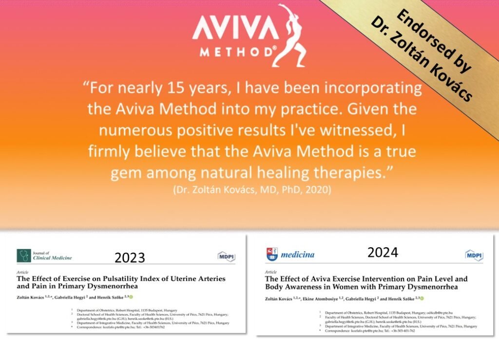
The Aviva Method and Menstrual Pain Relief
The Aviva Method is a structured system of targeted exercises developed to support female reproductive health. It has been practiced for nearly six decades and was recently examined in a large-scale, seven-year scientific research project conducted between 2018 and 2025.
Scientifically observed effects include:
immediate changes in uterine artery blood flow measured by ultrasound
significant reduction in menstrual pain after consistent practice across two menstrual cycles
improved body awareness, allowing earlier and gentler responses to discomfort
Research suggests that practicing the exercises at least twice per week is necessary to achieve these effects (Kovács et al., 2023; Kovács et al., 2024).
Aviva Exercises During Menstruation
Specific Aviva exercises are considered safe and supportive even during active bleeding, particularly for individuals experiencing painful menstruation.
These include the 4th and 6th exercises of the basic Aviva sequence (Steiner, 2009).
These movements may:
Gently mobilize the pelvis.
Improve the elasticity of pelvic blood vessels.
Support circulation in the lower abdomen
Assist the uterus in breaking down and expelling the endometrial lining and aggregated blood material more efficiently.
When resistance is reduced, the uterus can work with less force, which is often associated with decreased pain intensity.
Importantly, these exercises are not designed to elicit muscular effort. They aim to cooperate with physiological processes rather than override them.
A Body That Is Signaling, Not Failing
Menstrual pain is not something to suppress or ignore. It is often a signal pointing toward the need for improved circulation, reduced tension, and better nervous system support.
For many people, the Aviva Method offers a structured, movement-based, and evidence-informed approach to addressing these contributing factors.
If you would like to explore this further, you can learn more about the Aviva Method on my homepage at vitalityflame.com or participate in an online 2×2-hour Aviva Foundation session through Vitality Flame.
Sometimes, one carefully chosen movement is enough to begin changing the conversation between you and your body.
References
Ballantyne, J., Eisenberg, E., Giamberardino, M., Sharify, M., & Sjölund, B. (2008). Pain clinical updates. International Association for the Study of Pain.
Beller, F. (1971). Observations on the clotting of menstrual blood and clot formation. American Journal of Obstetrics and Gynecology, 109, 121–126.
Carroquino-García, P., Jiménez-Rejano, J., Medrano-Sánchez, E., de la Casa-Almeida, M., Díaz-Mohedo, E., & Fernández-de-Las-Peñas, C. (2019). Therapeutic exercise in the treatment of primary dysmenorrhea: A systematic review and meta-analysis. Physical Therapy, 99(10), 1371–1380.
Davies, J., & Kadir, R. (2012). Endometrial haemostasis and menstruation. Reviews in Endocrine & Metabolic Disorders, 13(4), 289–299.
Dawood, M. Y. (2006). Advances in pathogenesis and management of dysmenorrhea. Obstetrics and Gynecology, 108(2), 428–441.
Elovitz, M., Saunders, T., Ascher-Landsberg, J., & Mark, H. (2000). Effects of thrombin on myometrial contractions in vitro and in vivo. American Journal of Obstetrics and Gynecology, 183(4), 799–804.
Ferenczy, A. (2003). Pathophysiology of endometrial bleeding. Maturitas, 45(1), 1–14.
Hahn, L., Cederblad, G., Rybo, G., Pehrsson, N., & Korsan-Bengtsen, K. (1976). Blood coagulation, fibrinolysis and plasma proteins in women with normal and with excessive menstrual blood loss. British Journal of Obstetrics and Gynaecology, 83, 933–939.
Hickey, M., & Fraser, I. (2000). Clinical implications of disturbances of uterine vascular morphology and function. Baillière’s Best Practice & Research: Clinical Obstetrics & Gynaecology, 14(6), 937–951.
Kovács, Z., Atombosiye, E., Hegyi, G., & Szőke, H. (2024). The effect of Aviva exercise intervention on pain level and body awareness in women with primary dysmenorrhea. Medicina (Kaunas), 60(1), 184. https://doi.org/10.3390/medicina60010184
Kovács, Z., Hegyi, G., & Szőke, H. (2023). The effect of exercise on pulsatility index of uterine arteries and pain in primary dysmenorrhea. Journal of Clinical Medicine, 12(22), 7021. https://doi.org/10.3390/jcm12227021
László, K. D., Győrffy, Z., Ádám, S., Csoboth, M., & Kopp, M. S. (2008). Work-related stress factors and menstrual pain: A nationwide representative survey. Journal of Psychosomatic Obstetrics & Gynecology, 29(2), 133–138.
Liegard, W., & Brown, W. (1855). On uterine pains and hemorrhage after delivery. Atlanta Medical and Surgical Journal, 11, 241–248.
Núñez-Troconis, J., Carvallo, D., & Martínez-Núñez, E. (2021). Primary dysmenorrhea: Pathophysiology. Investigación Clínica, 62(2), 123–132.
Proctor, M., & Farquhar, C. (2006). Diagnosis and management of dysmenorrhoea. British Medical Journal, 332(7550), 1134–1138.
Rajput, V. (2021). An overview on dysmenorrhea. International Journal of Nursing Education and Research, 9(1), 1–4. https://doi.org/10.5958/2454-2660.2021.00001.X
Rosenwaks, Z., & Seegar-Jones, G. (1980). Menstrual pain: Its origin and pathogenesis. Journal of Reproductive Medicine, 25(4), 207–212.
Steier, A. (2009). Az AVIVA MÓDSZER: AVIVA’S METHOD S.O.S nőknek (Vol. I). Budapest. ISBN: 978-963-550-738-2 / 978-963-550-739-9
Vijayasri, R., Preetha, S., Abitha, P., Anushya, D., & Bhuvaneshwari, V. (2023). Dysmenorrhea decoded: Unveiling the mysteries of painful period. Journal of Gynecology and Women’s Health, 26(5). https://doi.org/10.19080/JGWH.2023.26.556183